 D.S. is a 56-year-old man with a two-year history of intermittent swelling of the right neck who was referred by his primary care physician for evaluation. He related that the swelling was mostly associated with eating, and would begin as soon as he started to eat. The swelling was painful and usually lasted several hours. He had not found anything that relieved it other than just waiting for it to go down.
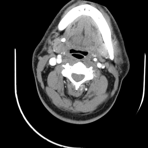
On examination, he was noted to have a firm right submandibular gland (SMG), but was otherwise normal. A CT scan was performed with findings as seen in image (right). There is a stone in the right SMG that measures about 6x7 mm. The stone is at the junction of the SMG duct and the hilum of the gland.
Because of his symptoms, it was recommended that he undergo sialendoscopy with attempted removal of the stone. We discussed that due to the relatively large size of the stone. it would likely be necessary to use the Holmium-Yag laser to break up the stone for removal, and if that was not possible, the gland should be removed.
He wished to proceed. and he underwent successful sialendoscopy with laser lithotripsy as is seen in the video. At his one-month follow-up appointment, he was doing well with resolution of his symptoms.at can be manually palpated can typically be removed trans-orally as in this case. The sialendoscope is helpful to trans-illuminate the duct and identify the exact location of the stone. For parotid stones, an external skin incision is required to use this approach.